4450
Population analysis of B0 magnetic field conditions in the human heart1Department of Biomedical Engineering, Columbia University, New York, NY, United States, 2Department of Radiology, Columbia University Irving Medical Center, New York, NY, United States, 3Section of Medical Physics, Department of Radiology, Mainz University Hospital, Mainz, Germany, 4Chair of Molecular and Cellular Imaging, Comprehensive Heart Failure Center (CHFC), Würzburg, Germany
Synopsis
Cardiac MRI suffers susceptibility-induced artifacts due to B0 inhomogeneity across the heart. The lack of population data in cardiac B0 conditions and the practical inability to obtain such data in large populations impedes the development of optimal cardiac B0 shim strategy. Here, we establish population-based B0 conditions from readily available CT images and simulate cardiac B0 maps of 254 CT subjects with broad demographic parameters. The results are expected to develop optimal subject- and population-specific cardiac B0 shim strategies.
Introduction
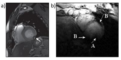
Cardiac MRI has been considered as a gold standard to access cardiac function1. Cardiac functional scans adopting balanced steady-state free precession (bSSFP) sequences at 3 T and multiecho gradient echo (GRE) sequences at 7 T suffer from susceptibility-induced artifacts2-6 (Figure 1) in the myocardium due to B0 variations across the heart. The best remedy to mitigate these issues is cardiac B0 shimming7, which requires in vivo B0 maps in the heart typically acquired with breath-hold8. However, the lack of population data in cardiac B0 conditions, especially for the patients with impaired lung capability9, in pediatrics10, and in elderly11 who are not able to undergo breath-hold for B0 acquisition, impedes the development of optimal cardiac B0 shim strategy. To overcome this challenge, we propose to investigate the cardiac B0 conditions in the population via B0 simulation from a large sample of CT images. This abstract presents the preliminary results of cardiac B0 maps from 254 subjects as part of an ongoing research study with an enrollment goal of 1000 subjects. These B0 maps were analyzed with spherical harmonic (SH) shimming. The resultant B0 conditions were investigated by computing their correlations associated with the subjects' demographic parameters. This study allows us to investigate the distributions and features of cardiac B0 conditions in population groups and pave the way to develop optimal cardiac B0 shim strategies.Methods
High-resolution CT images and demographic parameters of 254 adult subjects (Female: 136, Male: 118) who underwent a clinically indicated diagnostic CT scan were retrospectively collected and anonymized in accordance with our institutional review board requirements. The normality of subjects’ demographic parameters, including age, height, weight, body mass index (BMI), was tested using the Shapiro-Wilk test. Whole thoracic 3D CT images were down-sampled from 0.7-1.3 mm resolution to 1.5 mm isotropic spatial resolution consistent across all subjects. Then, the B0 distribution in the heart of each subject was calculated under the background field strength of 3 T and 7 T by an established B0 simulation approach12,13. The field calculation was performed in B0DETOX software14.B0 maps are typically decomposed into SH terms, and the coefficient of each term is scaled to the shim current of the corresponding shim coil in practical B0 shimming. To investigate the B0 conditions associated with the best possible shim for the whole heart, the simulated B0 maps of each subject were decomposed up to 2nd and 3rd SH order without constraints. Shim coils in MR scanners typically have output limits due to shim capability of the hardware. To investigate the impact of vendor-specific shim limits on the residual B0 inhomogeneity, we also performed the shim analysis up to 2nd SH order at 3 T with calibrated shim limits of scanners at Columbia University, including GE Premier, GE MR750, and Siemens Prisma. The shim analysis up to 3rd SH order was performed using the same limits of the 2nd order while leaving the 3rd order unlimited. Moreover, shim analysis at 7 T was performed with calibrated shim limits up to 3rd order from Siemens Terra at the University Hospital Würzburg (UKW), Germany. The resultant B0 inhomogeneity was presented as the standard deviation (σ) of the residual B0 distribution after removing corresponding SH shim terms.
To seek if population groups share specific aspects of cardiac B0 shapes, we calculated the correlation between unconstrained SH coefficients, B0 inhomogeneities at 3 T, and the height, weight individually. The B0 inhomogeneity between female and male groups was compared using the student’s t-test, and the significance criteria p<0.05 was adjusted using Bonferroni correction.
Results
Figure 2 shows the distribution of the subjects’ demographic parameters: age: 63±14 (Mean±SD) years (Shapiro-Wilk test: W=0.984, p-value<0.01), height: 1.67±0.11 m (W=0.987, p-value<0.05), weight 76.0±19.9 kg (W=0.965, p-value<0.001), BMI: 27.1±6.3 kg/m2 (W=0.967, p-value<0.001). The SH coefficients up to 2nd SH order and up to 3rd SH order were presented at 3 T (Figure 3) and 7 T (Figure 4). B0 inhomogeneity after unconstrained 2nd and 3rd shim at 3 T were 36±6 Hz and 27±5 Hz, respectively, while the corresponding results at 7 T were 83±13 Hz and 64±11 Hz. The shim capability of GE Premier showed limitation at 2nd order while Siemens Terra had limitations at 3rd SH order, especially for the Z3 term, leading to increased B0 inhomogeneity for some subjects after shim. The selected correlations between B0 conditions and demographic parameters were shown in Figure 5. Female subjects showed significantly lower B0 inhomogeneity than male subjects before and after 2nd/3rd shim. Z3 term and height have a maximum correlation of 0.343.Discussion
Here we present a detailed analysis of B0 conditions in the human heart from 254 subjects. The results suggest the 2nd and 3rd order SH shim requirements for the cardiac B0 shimming at 3 T and 7 T. The association between B0 conditions and demographic parameters allows us further understand the distribution and detailed characterization of B0 field conditions in the heart. Future research will involve more CT subjects, and the population analysis of cardiac B0 conditions is expected to enable optimal subject- and population-specific cardiac B0 shim strategies for clinical use cases.Acknowledgements
No acknowledgement found.References
1. Wieben O, Francois C, Reeder SB. Cardiac MRI of ischemic heart disease at 3 T: potential and challenges. Eur. J. Radiol. 2008;65(1):15-28.
2. Rajiah P, Bolen MA. Cardiovascular MR imaging at 3 T: opportunities, challenges, and solutions. Radiographics. 2014;34(6):1612-35.
3. Schär M, Kozerke S, Fischer SE, Boesiger P. Cardiac SSFP imaging at 3 Tesla. Magn. Reson. Med. 2004;51(4):799-806.
4. Meloni A, Hezel F, Positano V, Keilberg P, Pepe A, Lombardi M, Niendorf T. Detailing magnetic field strength dependence and segmental artifact distribution of myocardial effective transverse relaxation rate at 1.5, 3.0, and 7.0 T. Magn. Reson. Med. 2014;71(6):2224-2230.
5. Hock M, Terekhov M, Stefanescu MR, et al. B0 shimming of the human heart at 7T. Magn. Reson. Med. 2021;85(1):182-196.
6. Reiter T, Lohr D, Hock M, et al. On the way to routine cardiac MRI at 7 Tesla-a pilot study on consecutive 84 examinations. PLoS One. 2021;16(7):e0252797.
7. Kubach MR, Bornstedt A, Hombach V, Merkle N, Schär M, Spiess J, Nienhaus GU, Rasche V. Cardiac phase-specific shimming (CPSS) for SSFP MR cine imaging at 3 T. Phys. Med. Biol. 2009;54(20):N467.
8. Huelnhagen T, Hezel F, Serradas Duarte T, et al. Myocardial effective transverse relaxation time Correlates with left ventricular wall thickness: A 7.0 T MRI study. Magn. Reson. Med. 2017;77(6):2381-2389.
9. Pednekar AS, Wang H, Flamm S, Cheong BY, Muthupillai R. Two-center clinical validation and quantitative assessment of respiratory triggered retrospectively cardiac gated balanced-SSFP cine cardiovascular magnetic resonance imaging in adults. J. Cardiovasc. Magn. Reson. 2018;20(1):1-11.
10. Pednekar AS, Jadhav S, Noel C, Masand P. Free-breathing Cardiorespiratory Synchronized Cine MRI for Assessment of Left and Right Ventricular Volume and Function in Sedated Children and Adolescents with Impaired Breath-holding Capacity. Radiology: Cardiothoracic Imaging. 2019;1(2):e180027.
11. Kalva SP, Mueller PR. Vascular imaging in the elderly. Radiol. Clin. North Am. 2008;46(4):663-683.
12. Shang Y, Theilenberg S, Schreiber LM, Juchem C. Optimization of B0 Simulation Strategy in the Human Heart based on CT Images at limited Field of View. Proc Int Soc Magn Reson Med 2021:3635.
13. Shang Y, Theilenberg S, Mattar W, Terekhov M, Jambawalikar SR, Schreiber LM, Juchem C. High Resolution Simulation of B0 Field Conditions in the Human Heart Based on Segmented CT Images. Proc Int Soc Magn Reson Med 2019:2184.
14. Juchem C. B0DETOX - B0 Detoxification Software for Magnetic Field Shimming. Columbia TechVenture (CTV), License CU17326. 2017;innovation.columbia.edu/technologies/cu17326_b0detox
Figures


