2567
Classification of Gulf War Illness Patients vs Control Veterans Using fMRI Dynamic Functional Connectivity1Computer Engineering, University of Houston-Clear Lake, Houston, TX, United States, 2Emory University, Atlanta, GA, United States, 3University of Texas Southwestern Medical Canter at Dallas, Dallas, TX, United States
Synopsis
Around 200,000 veterans suffer from Gulf War Illness (GWI). GWI is characterized by multiple deficits in cognitive, emotion, somatosensory and pain domains. In this study we studied 23 GWI patients and 30 age-matched control veterans with resting-state fMRI in order to classify patients versus controls using dynamic functional connectivity among brain networks. Results show that different brain networks have discriminating power, pointing to widespread impairments in functional connectivity of visual, semantic, multi-sensory, and sensory-motor processing networks in GWI, consistent with multi-symptom nature of GWI.
INTRODUCTION
Gulf War Illness (GWI) is a chronic medical condition characterized by multiple symptoms which indicate brain function deficits in cognitive, pain, emotion, and somatosensory domains1-3. It affects approximately 200,000 of the 1991 Gulf War veterans. Prior neuroimaging studies confirmed presence of structural, functional and metabolic brain impairments in GWI4-7; however, GWI is still poorly understood. During the last three decades, functional neuroimaging technology, especially functional magnetic resonance imaging (fMRI), has improved tremendously, with recent attention towards resting-state dynamic functional connectivity (DFC) analysis of the brain8-14. DFC analyses can provide insight into overall functional connectivity between brain networks; differences in the DFC of brain networks under different brain conditions can be studied and used as features for classification14. In this study we use standard deviation of the DFC fluctuations (stdDFC) among brain regions as features to classify the GWI patients from normal control (NC) veterans, and compare the results with those of static FC.METHODS AND MATERIALS
23 GWI veterans (mean age 49.4 yrs.) and 30 normal control (NC) veterans (mean age 49.8 yrs.) were scanned in a Siemens 3T Tim Trio MRI scanner using a 12-channel receiver head coil. Written informed consent was obtained from all participants in the protocol approved by the local Institutional Review Board. Wholebrain resting-state fMRI (rsfMRI) data were acquired with a 10-min whole-brain gradient echo EPI (TR/TE/FA = 2000/24ms/90°, resolution = 3mm×3mm×3.5mm). Resting-state fMRI pre-processing steps included attenuation of signal related to subject-motion and physiological responses, using advanced ICA-based artifact reduction techniques, and spatial smoothing with FWHM = 6mm isotropic Gaussian kernel. Spatially-averaged fMRI signals were obtained for each of the regions using a 116-region AAL Atlas15. Static FC and DFC were computed for each of the Combination(116-of-2) = 6670 region-pair combinations. For the DFC, the standard deviation (std) of the DFC fluctuations across the time-windows (i.e. std of the temporal fluctuations/dynamics of the FC, or stdDFC) was computed for each pair, and the stdDFCs were used as features. For the static FC, the FC value for each region-pair was used as features. To reduce the number of features, unpaired t-test was done to find the most significantly different region-pairs across the two groups, under different p-value thresholds ranging from 0.1 to 0.001. These reduced set of features were used in classification with support-vector machine (SVM) classifier, using 10-fold cross-validation and 100 iterations. Average value of the classification accuracy (ACA) across the iterations was computed.RESULTS
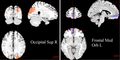
Classification using the DFC-based method resulted in ACA of as high as 98%, for the threshold of p<0.05, resulting in 113 region-pairs. ACA of 94%, 81% and 65% were achieved by using only 32 pairs (p<0.02), 12 pairs (p<0.01), and a single pair (p<0.001), respectively. ACA decreased when more than 113 pairs were used (i.e. p>0.05). The region-pair that appeared consistently in all of these above region-pair sets was the pair consisting of AAL region #25, Left Medial Orbital Superior Frontal Gyrus, and, region #50, Right Superior Occipital Gyrus. These two regions are presented as overlays on a standard individual T1w MRI image in Figure 1. The average value of the stdDFC for the GWI group was 0.226, which was significantly lower (p<0.001) than that of the NC group, which was 0.272.For comparison, the (static) FC-based classification, the highest ACA was 72% using 13 region-pairs (p<0.05). Using various thresholds of p values ranging from 0.001 to 0.1 resulted in 1 to 480 significant region-pairs, which resulted in 66% to 72% ACA, using all pairs further decreased ACA. The region-pair that appeared consistently in all of these (static) FC pairs was the pair consisting of AAL region #74, Right Putamen, and region #116, Vermis10.
DISCUSSIONS AND CONCLUSIONS
Dynamic functional connectivity between multiple brain networks appeared as having good group discriminating power with an average classification accuracy of up to 98%, whereas static FC-based method achieved at most 72% accuracy. It is also interesting to note that, the range of the fluctuations in the DFC between the Right Superior Occipital Gyrus and the Left Medial Orbital Superior Frontal Gyrus, regions during the resting-state fMRI scan, as captured by stdDFC, which was 17% lower for the GWI than the stdDFC of the NC group (significantly lower, with p<0.001). This result may potentially signal an impairment for the GWI group between these regions, which are involved in functions such as visual processing, multi-sensory input processing sensori-motor processing and semantic processing. Consistent with these findings, GWI veterans were reported to exhibit deficits in word-finding6, in visual processing16, and in fine motor skills3. However, brain networks involved in successful classification need to be further interpreted and studied. Ongoing and future work involves different feature selection and classification algorithms to achieve a higher classification accuracy, as well as more detailed study of other region-pairs involved in group discrimination. Overall, the results are in line with other recent findings of widespread impairments in resting-state FC within brain function networks implicated by multiple symptoms in GWI patients1-7. DFC-based metrics, such as the stdDFC in our study, with their group-discriminating differences can potentially lead to resting-state fMRI / neuroimaging biomarkers for GWI, and also potentially for other neurological disorders and conditions.Acknowledgements
This work was supported by the Office of Assistant Secretary of Defense for Health Affairs, through the Gulf War Illness Research Program under Award No.W81XWH-16-1-0744 (PI: Gopinath). Opinions, interpretations, conclusions and recommendations are those of the authors and are not necessarily endorsed by the Department of Defense.References
[1] Haley RH, et al., JAMA, 277: 215–222 (1997).
[2] Toomey R, etal., J Int Neuropsychol Soc., 15:717–729 (2009).
[3] Binns JH, et al., Report of Research Advisory Committee on Gulf War Veterans’ Illnesses, In:Affairs DoV, editor, Boston, MA, U.S. Government Printing Office (2014).
[4] Li X, et al., Radiology, 261:218–225 (2011).
[5] Gopinath K, et al., Neurotoxicology, 33:261–271 (2012).
[6] Moffet K, et al., Brain Cogn., 98:65–73 (2015).
[7] Gopinath K, et al., Neuroscience Letters, 701:136–141 (2019).
[8] Sakoglu U, et al., NeuroImage, 47 (Suppl. 1):S57 (2009).
[9] Sakoglu U, et al., NeuroImage, 47 (Suppl. 1):S169 (2009).
[10] Sakoglu U, et al., MAGMA, 23(5-6):351–366 (2010).
[11] Chang C, et al., NeuroImage, 50(1):81–98 (2010).
[12] Hutchison RM, et al., NeuroImage, 80(1):360–378 (2013).
[13] Allen E, et al., Cereb Cortex, 24(3): 663–676.80 (2014).
[14] Sakoglu et al., J Neurosci Res., 97(7):790–803 (2019).
[15] Automated Anatomical Labeling (AAL) Atlas, https://www.gin.cnrs.fr/en/tools/aal.
[16] White RF, et al. Am J Ind Med., 40:42–54 (2001).
Figures
