0453
Simultaneous Multislice pTx for Readout-Segmented Diffusion Imaging at 7 T1Imaging Centre of Excellence, University of Glasgow, Glasgow, United Kingdom, 2Siemens Healthcare Ltd. United Kingdom, Frimley, United Kingdom, 3Siemens Healthcare GmbH, Erlangen, Germany
Synopsis
This abstract addresses the challenges of 7T diffusion MRI associated with shorter T2/T2* and inhomogeneous B1+ field by implementing simultaneous multislice (SMS) parallel-transmit (pTx) slice-by-slice shimming in a readout-segmented diffusion-weighted sequence, RESOLVE. The benefits of pTx were compared to conventional single-transmit circularly polarized (CP) mode both with and without SMS using a matched high-resolution diffusion protocol in the same healthy volunteer. Slice-by-slice shimming significantly improves signal homogeneity while SMS opens the door for improved scan efficiency. The simplicity of slice-by-slice shimming and sequence integration makes this a practical solution for routine 7T imaging.
Introduction
The increase in SNR at 7T has been widely exploited to improve spatial resolution and image quality in a number of applications. However, it remains a challenge to realize these benefits with diffusion MRI (dMRI) due to the challenges of reduced T2 and T2* decay times, and the increased SAR. The shortened T2 leads to an increase in signal attenuation at typical TE values, whilst the reduced T2* limits the spatial resolution that can be achieved using single-shot EPI. The increased SAR can limit scan efficiency due to increased TR or further compound the T2 losses by increasing the duration of RF pulses. A further challenge within SAR constraints is the joint requirement for simultaneous multislice imaging (SMS) [1] to optimize scan efficiency, and parallel-transmit (pTx) [2,3] to mitigate B1+ field inhomogeneity at 7T. This study addresses some of these issues by combining the reduced TE and readout time of multishot EPI [4] with a static slice-by-slice B1+ shimming algorithm [5] that has been modified to accommodate multiband RF pulses for SMS [6].Methods
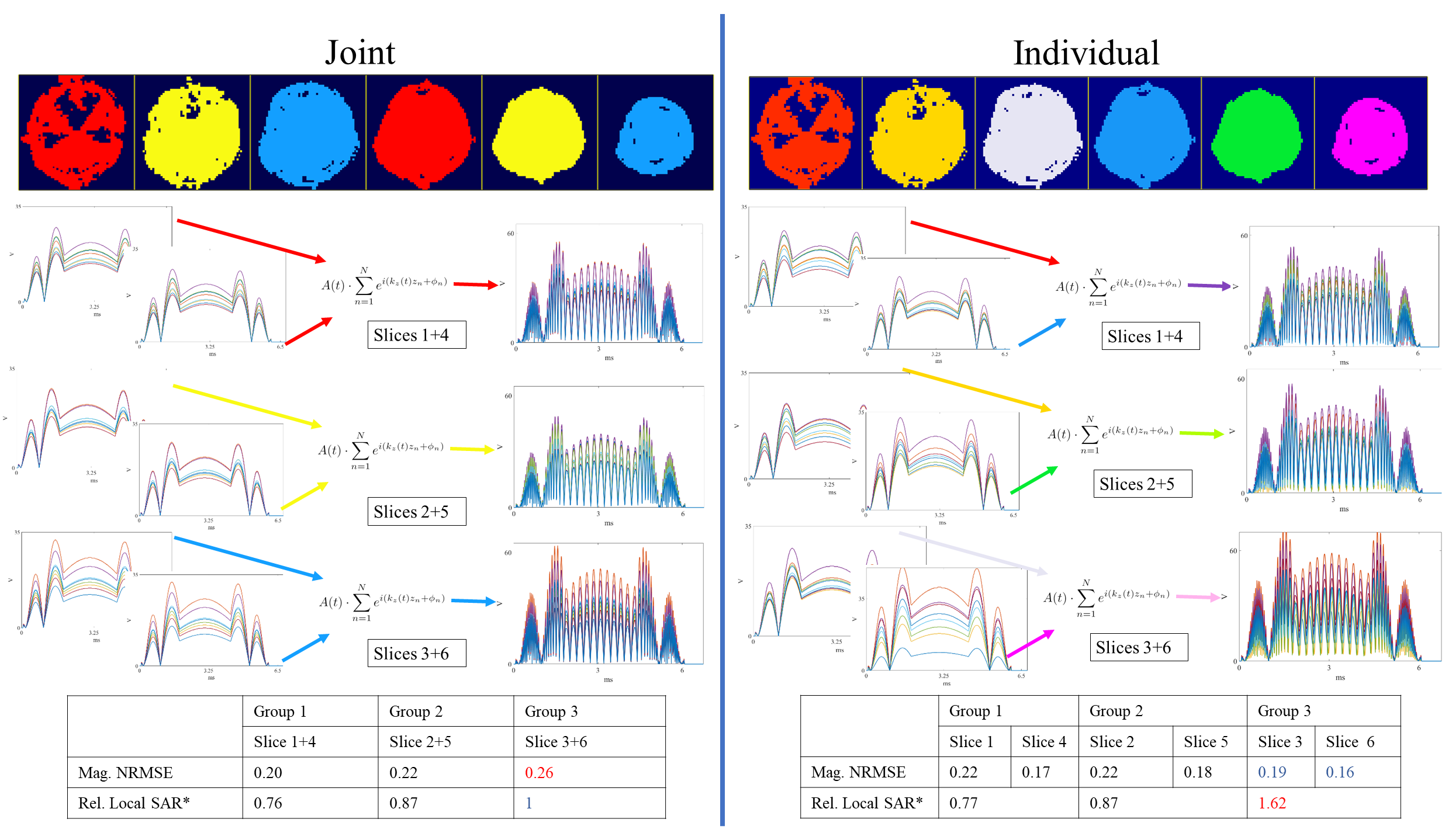
Data were acquired on a MAGNETOM Terra 7T Scanner (Siemens Healthineers, Erlangen, Germany) using a 2D readout-segmented, diffusion-weighted EPI sequence with a modification to support slice-selective pTx excitation and refocusing pulses with B1+ shimming. These pulses included conventional single-band (SB) excitation as well as SMS or multiband (MB) excitation with slice-GRAPPA reconstruction [1]. The pTx waveform envelopes were matched to the single-transmit equivalent sequence using circularly polarized (CP) excitation, which for SB was a conventional sinc waveform and for MB was a VERSE’d [7] version. The sequence navigator refocusing pulse [4] was maintained as CP.For SMS B1+ shimming, two optimization methods were explored: one in which shims were designed for each slice individually, and one in which shims were optimized jointly across the group of slices to be excited simultaneously (explained further in Figure 1). For both approaches, shims for excitation and refocusing were optimized in MATLAB’s $$$\texttt{fmincon}$$$ (Mathworks, MA, USA) using magnitude least-squares [8] with added nonlinear constraints on the standard deviation of the flip angle (≤25°) and a peak voltage limit of Vmax/MB, where Vmax was set to 180 V and MB refers to the number of slices excited simultaneously. After calculating B1+ shims for each slice (individually or jointly), the SMS pTx waveform was generated by summing and frequency shifting using the slice-gradient transmit k-space trajectory (Equation 2 Ref [9]). Optimal phase scheduling [10] was applied for MB=3. B1+ shim calculation time was negligible (<1 sec/slice) on a standard laptop (Intel(R)Core(TM)i5-8250U CPU).
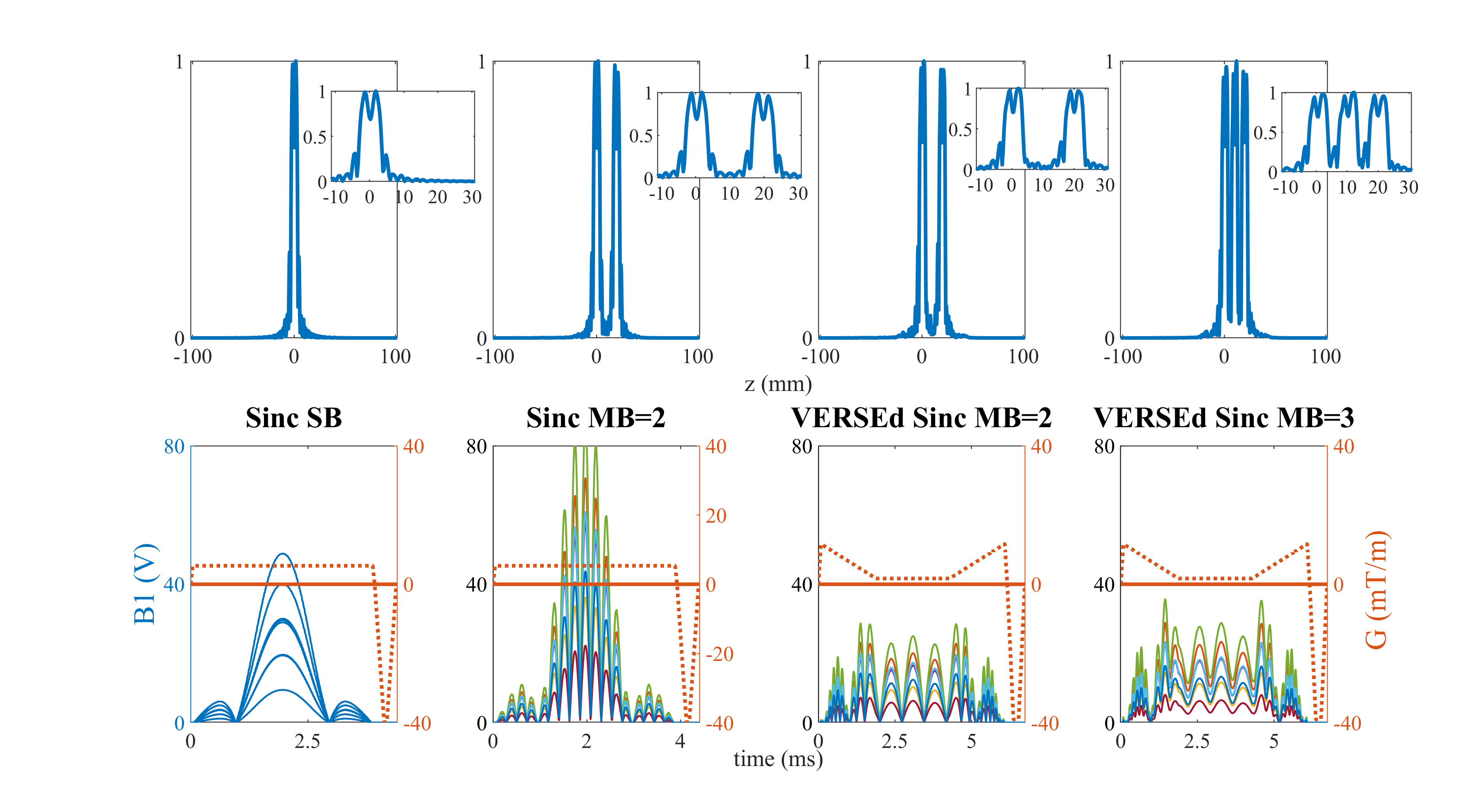
After verification of pTx SMS slice positioning and profiles in a phantom (Figure 2), a healthy volunteer was scanned in a locally-built 8Tx/32Rx head coil [11] following informed consent and local ethics committee approval. To compare the effects of CP vs pTx and furthermore SB vs MB, a time-matched protocol was used: TE1,SB/TE2,SB/TE1,MB/TE2,MB/TR/TA/RO segments/ESP/FOV/Slice Thickness/MAT/Slices/GRAPPA=58 ms/99 ms/69 ms/116 ms/2000 ms/4:35 min/11/0.32 ms/230 mm2/3 mm/306x306/6/3(2 for MB=3). Diffusion parameters: tetrahedral encoding (4-scan trace), b=0,1000 s/mm2 (2 averages each). For pTx, SB and MB=2 and 3 waveforms were generated using the joint design method (Figure 1). However, for the same pulse durations MB=3 was not possible with CP acquisition. Fat suppression was performed using frequency-selective saturation without gradient reversal.
Results
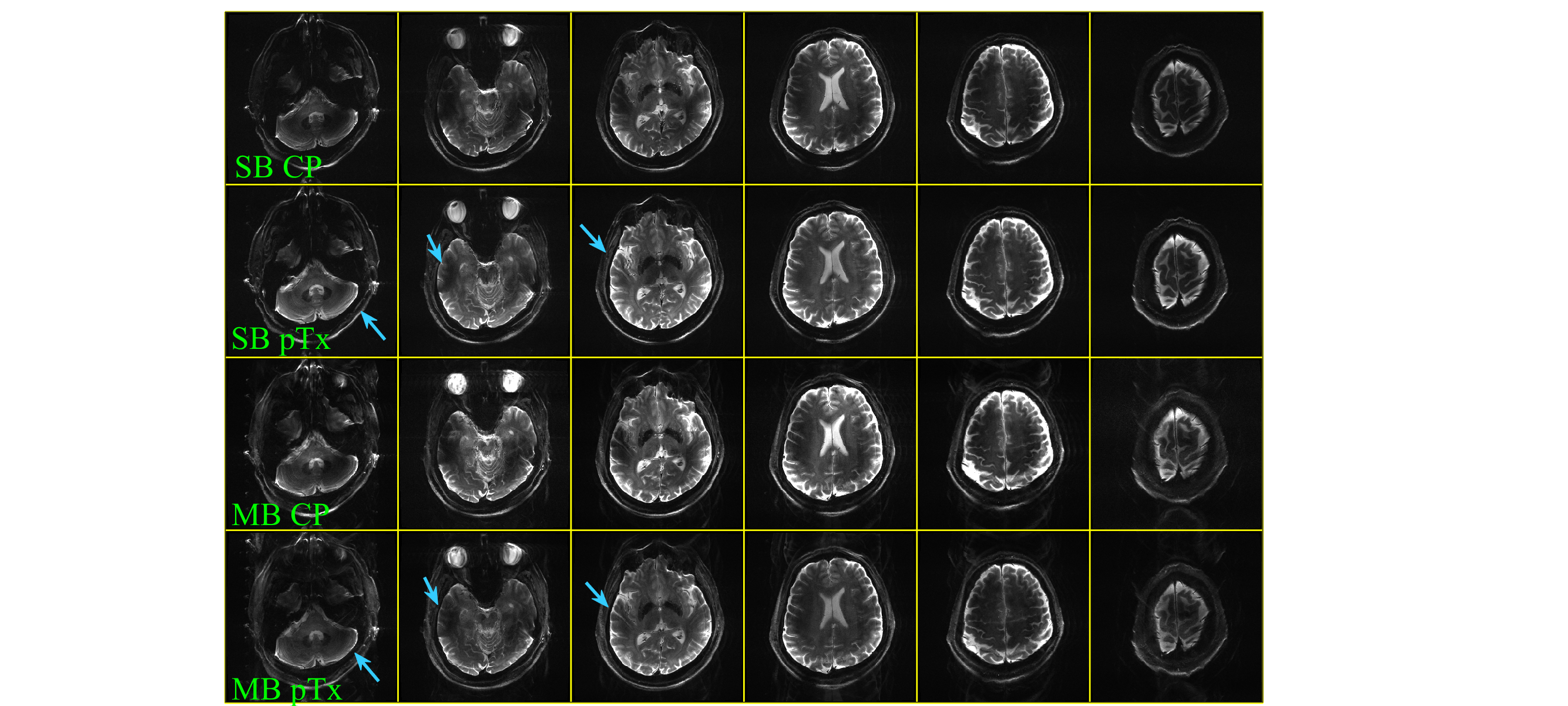
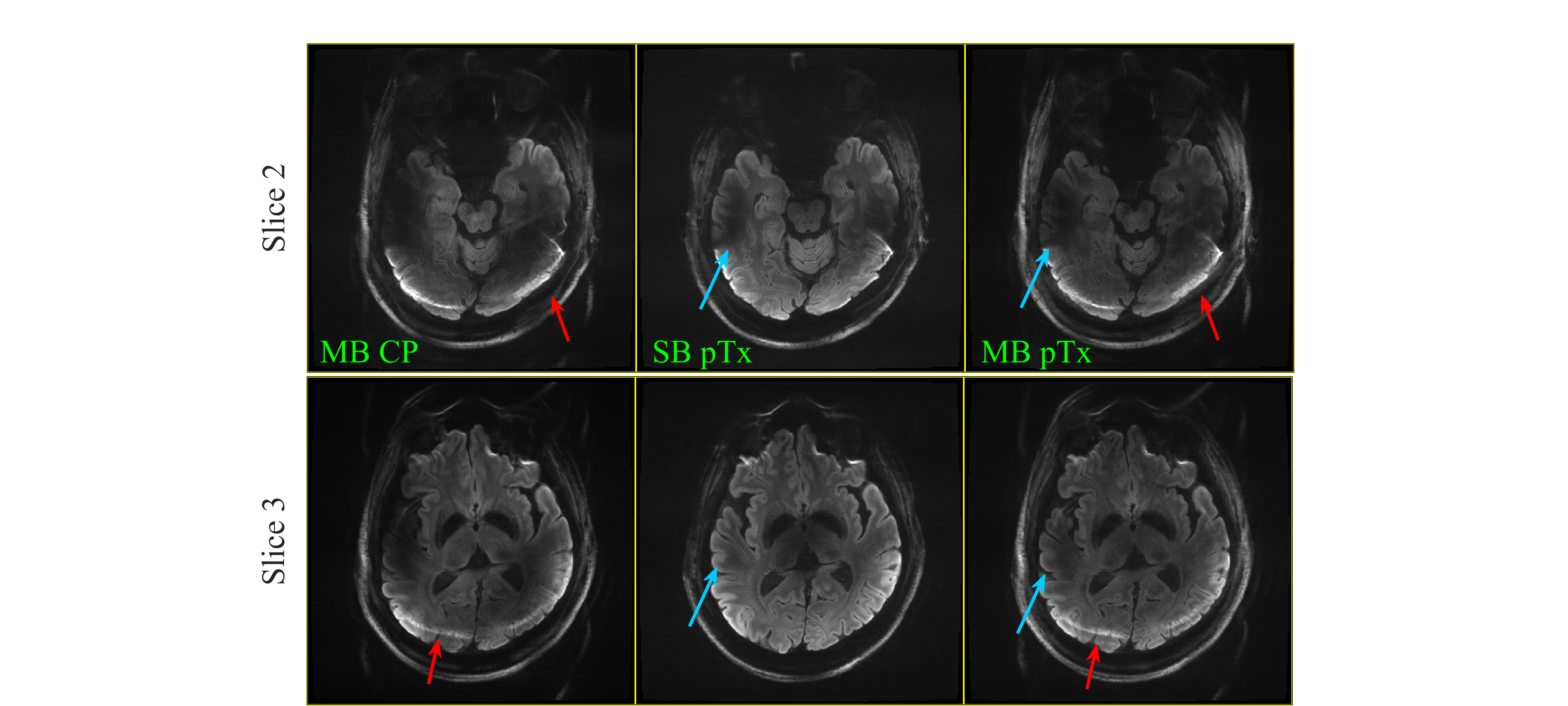
Figure 3 compares the b=0 s/mm2 images for CP and pTx (with slice-by-slice shimming) for both SB and MB excitation modes. An improvement in homogeneity is seen with pTx for both SB and MB acquisitions.Figure 4 shows trace-weighted b=1000 s/mm2 images at two slice positions for MB=2 in CP and pTx with slice-by-slice shimming. All MB images have an increased level of residual fat signal superimposed on tissue at the back of the brain due to poor fat suppression with both CP and pTx scans.
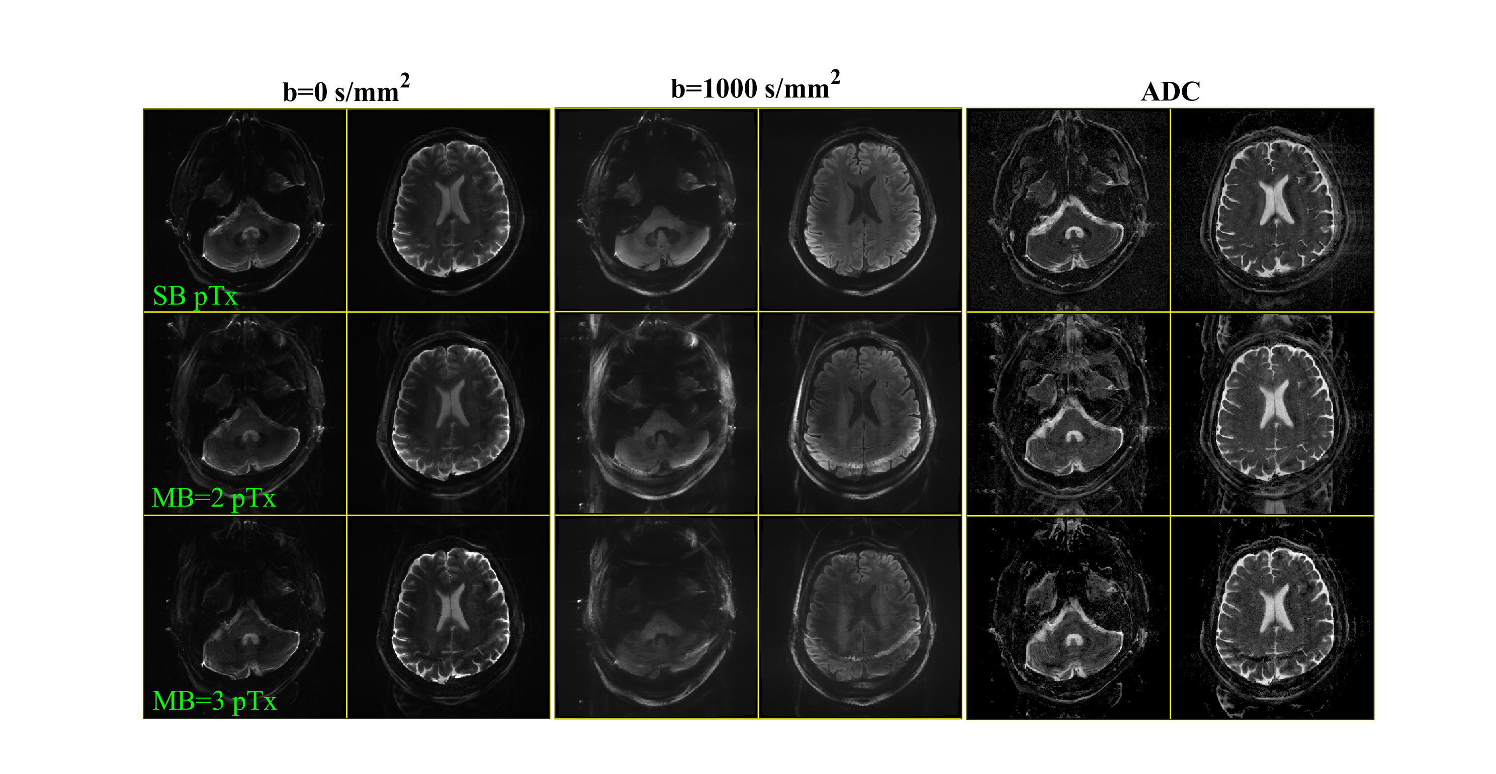
Figure 5 shows T2w, diffusion-weighted, and ADC images for pTx shimming with SB and MB=2 and 3 acquisitions. MB=3 was not possible for CP with the same VERSE pulse length due to peak voltage restrictions of the 180° refocusing pulse.
Discussion & Conclusion
In this work we present the implementation of pTx SMS within a commercially-available readout-segmented diffusion sequence, RESOLVE. Slice-by-slice shimming substantially improved the signal homogeneity without significant pulse design burden, and in conjunction with pTx SMS has the potential to permit scan acceleration by reducing TR (subject to SAR constraints and not explored in this preliminary work).pTx local SAR calculations via VOPs [12] make sequence preparation more computationally demanding at the scanner and is not yet incorporated into the pulse optimization. This currently limits the maximum slice number for SMS pTx. To avoid signal loss above the frontal sinuses, fat suppression was performed in this study without gradient reversal. However, this introduced a high level of chemical shift artifacts in both CP and pTx SMS images, which will need to be addressed in future work.
In conclusion, we have presented a potential solution for increasing scan efficiency and signal homogeneity in multishot diffusion-weighted imaging at 7T by incorporating SMS pTx. The proposed method is based on a straightforward B1+ shimming approach, which could be easily incoporated into routine 7T imaging.
Acknowledgements
Shajan Gunamony, Sarah Allwood-Spiers, Paul McElhinney, Rosemary Woodward, Tracey Hopkins, Evonne McLennan, Laura Dymock for their help and support scanning throughout the involved pTx development process.References
[1] K. Setsompop et al., “Blipped-Controlled Aliasing in Parallel Imaging for Simultaneous Multislice Echo Planar Imaging with Reduced g-Factor Penalty,” MRM, vol. 67, no. 5, pp. 1210-1224, Mar. 2012, doi: DOI 10.1002/mrm.23097.
[2] T. S. Ibrahim, R. Lee, B. A. Baertlein, A. Kangarlu, and P.-M. L. Robitaille, “Application of Finite Difference Time Domain Method for the Design of Birdcage RF Head Coils Using Multi-Port Excitations,” MRI, vol. 18, no. 6, pp. 733–742, Jul. 2000, doi: 10.1016/S0730-725X(00)00143-0.
[3] U. Katscher, P. Börnert, C. Leussler, and J. S. van den Brink, “Transmit SENSE,” MRM, vol. 49, no. 1, pp. 144–150, Jan. 2003, doi: 10.1002/mrm.10353.
[4] R. M. Heidemann, D. A. Porter, A. Anwander, T. Feiweier, K. Heberlein, T. R. Knösche, R. Turner. “Diffusion imaging in humans at 7T using readout-segmented EPI and GRAPPA”. Magn Reson Med. 2010 Jul;64(1):9-14. doi: 10.1002/mrm.22480.
[5] S. N. Williams, I. Dragonu, P. Liebig, and D. A. Porter, “Multi-Slice 2D pTx Readout-Segmented Diffusion-Weighted Imaging Using Slice-by-Slice B1+ Shimming,” in International Society for Magnetic Resonance in Medicine, Virtual, May 2021.
[6] X. Wu et al., “High-resolution whole-brain diffusion MRI at 7T using radiofrequency parallel transmission,” MRM, vol. 80, no. , pp. 1857-1870, Mar. 2018, doi: 10.1002/mrm.27189.
[7] S. Conolly, D. G. Nishimura, A. Macovski, and G. H. Glover, “Variable-Rate Selective Excitation,” JMR, vol. 78, pp. 440–458, Jan. 1988.
[8] K. Setsompop, L. L. Wald, V. Alagappan, B. A. Gagoski, and E. Adalsteinsson, “Magnitude Least Squares Optimization for Parallel Radio Frequency Excitation Design Demonstrated at 7 Tesla with Eight Channels,” MRM, vol. 59, no. 4, pp. 908–915, Apr. 2008, doi: 10.1002/mrm.21513.
[9] S. Abo Seada, A. N. Price, T. Schneider, J. V. Hajnal, and S. J. Malik, “Multiband RF Pulse Design for Realistic Gradient Performance,” MRM, vol. 81, no. 1, pp. 362–376, Jan. 2019, doi: 10.1002/mrm.27411.
[10] E. Wong, “Optimized Phase Schedules for Minimizing Peak RF Power in Simultaneous Multi-Slice RF Excitation Pulses,” in International Society of Magnetic Resonance in Medicine, Melbourne, Australia, May 2012.
[11] S. N. Williams et al., “A Nested Eight-Channel Transmit Array With Open-Face Concept for Human Brain Imaging at 7 Tesla,” Front. Phys., vol. 9, p. 701330, Jul. 2021, doi: 10.3389/fphy.2021.701330.
[12] G. Eichfelder and M. Gebhardt, “Local Specific Absorption Rate Control for Parallel Transmission by Virtual Observation Points,” MRM, vol. 66, no. 5, pp. 1468–1476, Nov. 2011, doi: 10.1002/mrm.22927.
Figures