3827
A Paradigm shift in MR physics with Deep Learning Reconstruction: higher image quality and spatial resolution in shorter scan time1Canon Medical System USA, Tustin, CA, United States
Synopsis
In MR physics, there is a fundamental tradeoff between image spatial resolution, signal to noise ratio, and scan time. To acquire images with high resolution and SNR, signal averaging is the most common solution, but results in longer scan time. In this study, a Deep Learning Reconstruction method was employed to remove the noise from clinical images and improve SNR. This SNR improvement was devoted to increase the spatial resolution without the need of signal averaging and increased scan time. Hence, higher resolution images with high image quality can be obtained in shorter time in clinical practice.
Introduction
In MR physics, there is a tradeoff between spatial resolution, Signal-to-Noise Ratio (SNR), and acquisition time. Increasing spatial resolution is desirable as it helps the clinicians visualize and identify small anatomical structures and pathologies. Higher spatial resolution negatively impacts SNR as there is less signal in a smaller voxel. Typically, to achieve high resolution without SNR drop, the acquisition is repeated multiple times and averaged. This repetition increases the scan time leading to patient discomfort and potential image artifacts due to patient motion.With Artificial Intelligence (AI) evolution, Deep Learning Reconstruction (DLR) methods are starting to play an essential role in MR imaging. Recently, commercial DLRs have been used to achieve higher resolution and SNR images1,2. By selectively removing the noise, the DLR improves the SNR without repeating the acquisition. Increasing SNR can be utilized in different ways: it can be used to improve the image resolution for better diagnosis; or to reduce the number of signal acquisition (NAQ) to achieve shorter scan time; or a combination of both. In this study, the DLR was used in multiple anatomical regions to achieve high spatial resolution in shorter scan time while preserving SNR and image contrast.
Methods
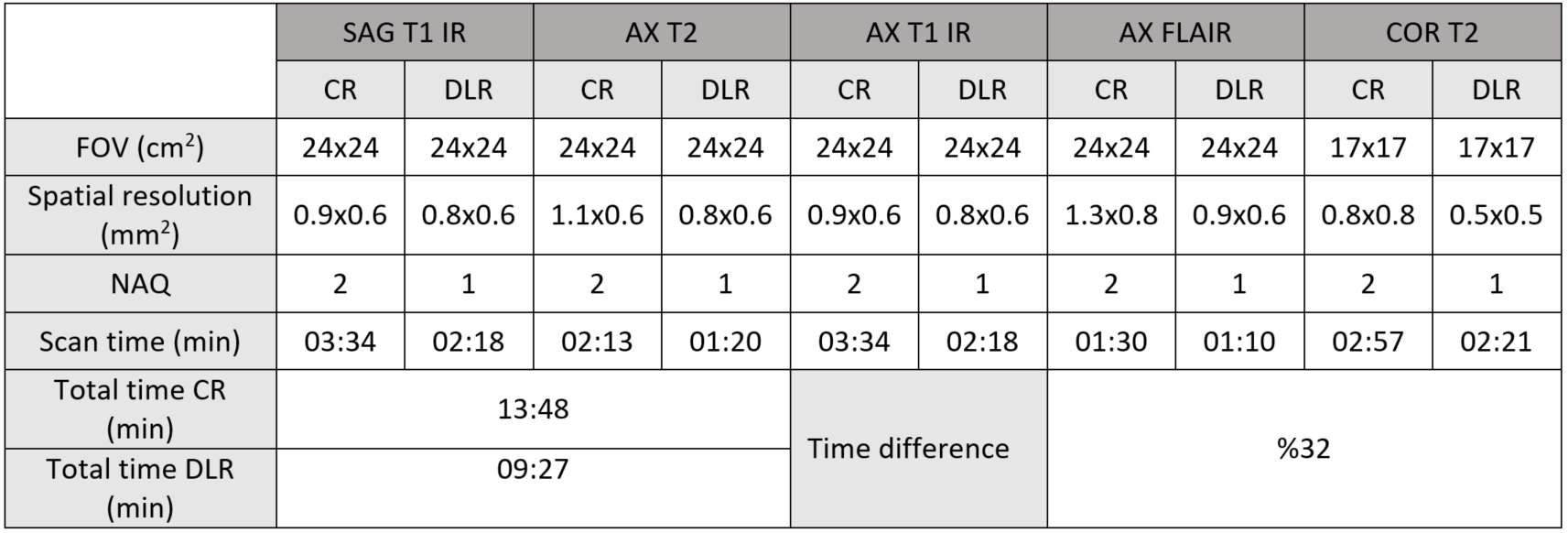
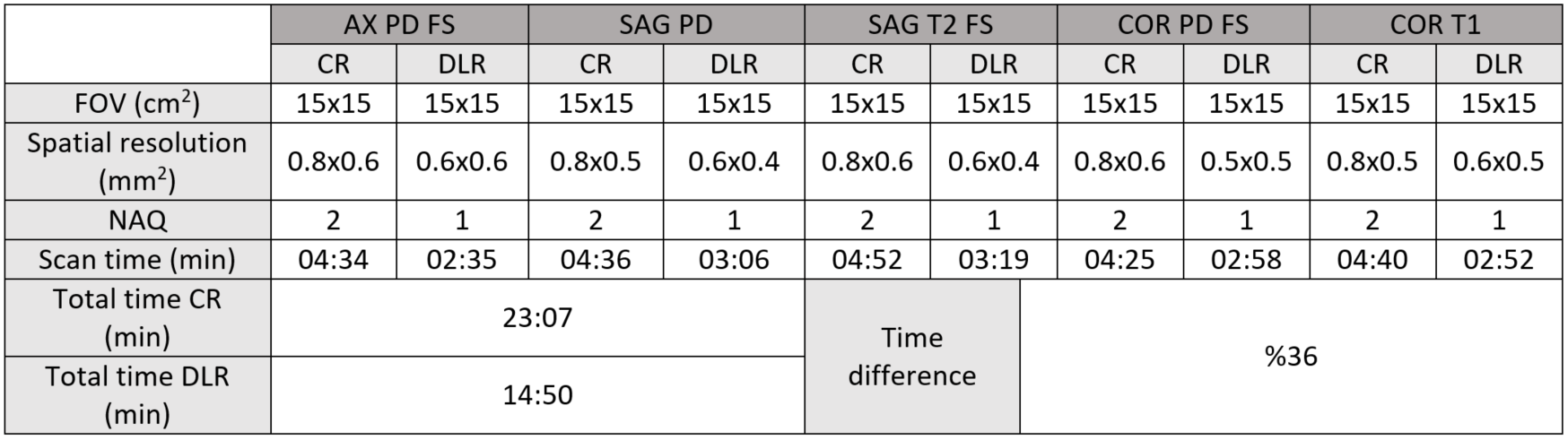
Routine clinical brain and knee scans were performed on volunteers on a Canon Vantage Orian 1.5T scanner. The scans were acquired using two protocols: a routine clinical protocol with a conventional reconstruction (CR) method using typical clinical post-processing filters to provide images with optimal smoothness and sharpness at the same time, and a modified protocol to achieve higher spatial resolution in a shorter scan time using the DLR method. Most sequence parameters were kept identical between the CR and DLR protocols. Wherever the DLR scan had an abundant SNR, spatial resolution was increased, and the NAQ was reduced in order to shorten the scan time. The altering parameters between two the protocols for each anatomical region and clinical sequence are shown in tables 1 and 2.To evaluate the impact of the DLR on image quality in brain, the SNR was measured in White Matter (WM) and Gray Matter (GM) and the contrast change between WM and GM was calculated in axial T1, T2, and FLAIR scans. Similarly, in knee, the SNR was measured in bone and muscle and the contrast change between bone and muscle was calculated for each sequence.
Results
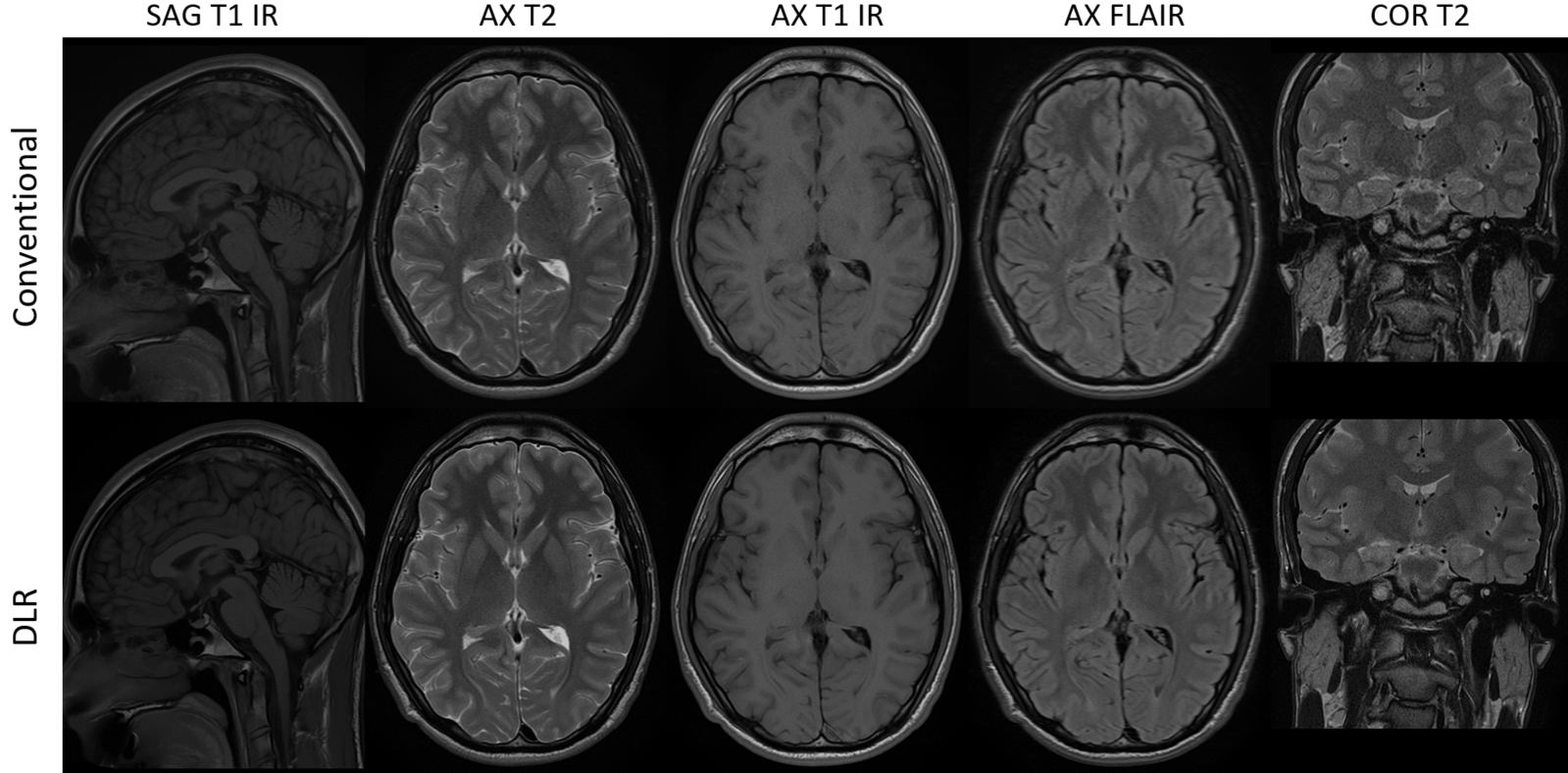
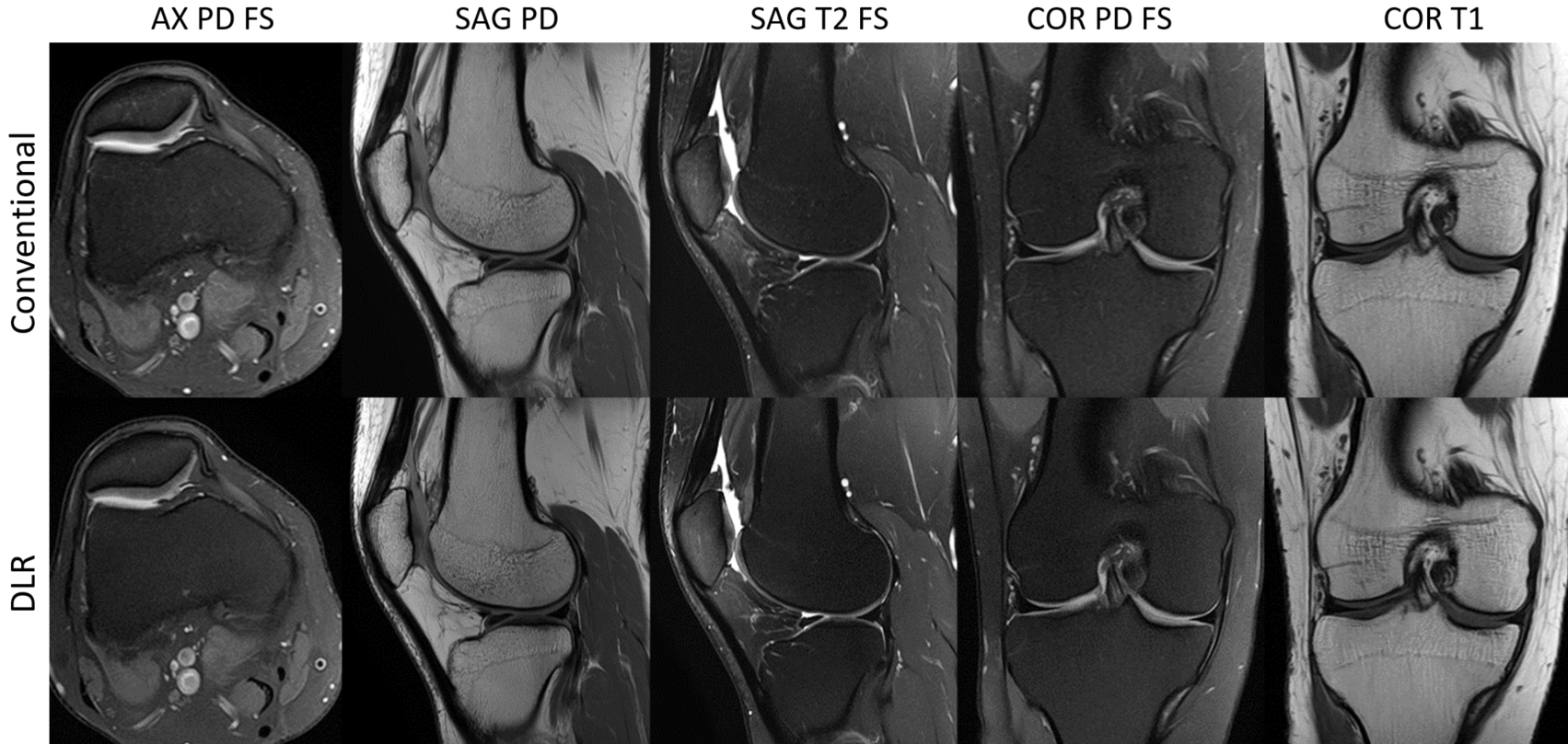
Figure 1 demonstrates a brain case acquired with the CR and DLR methods. As shown in table 1, the spatial resolutions in the DLR sequences were increased up to 37%. The scan time for each sequence was shorter than the CR sequences by reducing the NAQ in the DLR sequences, even though resolution was greater. The overall exam time was reduced 31.5% using DLR scans. The SNR was measured in GM and WM in several ROIs and averaged for each tissue type. As shown in table 3, the SNR using the DLR scan with higher resolution and shorter scan time was increased for all image contrasts. The contrast change between GM and WM was less than 3% for all sequences.Similar to the brain case, figure 2 shows a knee case where the spatial resolution was improved in DLR scan while the scan time for each sequence was reduced using only one NAQ. The overall scan time using the DLR was reduced 36% compared to the CR method. The measured SNR averaged in selected ROIs in femoral bone and muscle remained very similar between the CR and DLR scans in axial PD and coronal T2 scans while it slightly improved in bone in sagittal PD using the DLR scan. The contrast change between bone and muscle was less than 8% between the CR and DLR scans.
Discussions
As shown in figures 1 and 2, the DLR based images achieve a similar image quality to the CR images. In the brain scan, the spatial resolution of sub-millimeter was achieved with the DLR scan which resulted in sharper images. The higher resolution images were obtained while the SNR was improved and the scan time was reduced, contrary to MR Physics and conventional MR imaging techniques. The total routine brain scan with high resolution images was completed in less than 10 minutes. This can be very beneficial for clinical diagnosis as the shorter total scan time can provide more patient comfort and reduce patient motion and artifact. In addition, similar image quality with even higher resolution could improve the detection of small lesions in the brain.Similarly, in the knee scan, the spatial resolution was improved with DLR in all the sequences while the image quality, SNR and image contrast are similar to the CR scans. The time saving for the entire exam was 36% which can be advantageous for the musculoskeletal imaging wherein patients, usually experiencing severe pain, could benefit from the shorter scan time.
Conclusion
DLR facilitates higher resolution imaging in a shorter scan time while the clinical image quality is not compromised. The higher resolution with DLR scans results in sharper images which can be helpful to identify smaller structural details in the images and potentially lead to more reliable diagnosis and improved patient care.Acknowledgements
No acknowledgement found.References
1 Kidoh M, et al. Deep Learning Based Noise Reduction for Brain MR Imaging: Tests on Phantoms and Healthy Volunteers. Magn Reson Med Sci. 2019; doi:10.2463/mrms.mp.20190018.
2 Argentieri E, et al. Performance of a Deep Learning-Based MR Reconstruction Algorithm for the Evaluation of Peripheral Nerves. In Radiological Society of North America. 2019 Scientific Assembly and Annual Meeting; 2019.
Figures