1299
White Matter fiber orientation dependent R2*: comparison between post mortem in situ and in vivo1Institute of Forensic Medicine, Department of Biomedical Engineering, Basel, Switzerland, 2Institute of Forensic Medicine, Health Department Basel-Stadt, Basel, Switzerland, 3Department of Neuroradiology, Medical University of Innsbruck, Austria
Synopsis
White matter R2* imaging is sensitive to the fiber orientation with respect to the main magnetic field. This study compared fiber orientation dependent R2* of white matter in post mortem (pm) in situ and in vivo based on an identical technical approach using diffusion tensor imaging to estimate the fiber angle. R2* increased with increasing fiber angle in both groups, whereby a decreased R2* orientation sensitivity was observed post mortem compared to in vivo. This may be mainly caused by the decreased pm brain temperature compared to in vivo conditions leading to reduced diffusion and by tissue decomposition after death.
Introduction
R2* imaging of brain white matter (WM) is well known for being sensitive not only to myelin and iron content, but also to the orientation of nerve fibers with respect to the main magnetic field of the MRI scanner.1-4 The orientation dependency may be attributed to the anisotropic molecular environment in the myelin sheath as well as to inhomogeneities of the magnetic field due to the structural anisotropy of WM fiber tracts.5,6 The goal of the present work was to investigate the difference between post mortem in situ and in vivo WM R2* orientation dependency.Methods
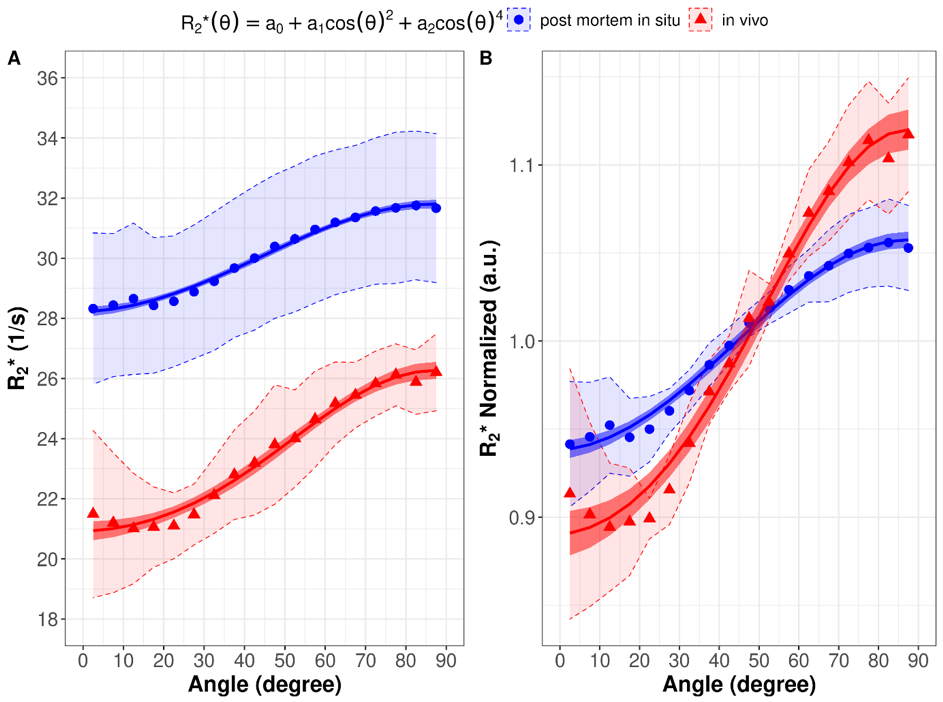
12 deceased subjects were examined and stored in a cooling chamber at 4° Celsius prior to study procedures. The in situ brain temperature was measured transethmoidally. In addition to the post mortem examinations, four healthy subjects volunteered for in vivo MRI. In order to determine the R2* orientation dependency, post mortem in situ and in vivo MRI scans were conducted at 3T. R2* was acquired with a multi-echo gradient echo sequence [12 TEs = 5.79-50.94 ms, TR = 68 ms, 40 slices, slice thickness 4 mm, in-plane resolution 1x1 mm2 and subsequent fitting of a mono-exponential decay signal model. The orientation of white matter fibers were computed using diffusion-weighted single-shot echo-planar imaging [64 isotropically distributed diffusion directions, TE = 109 ms, TR = 18700 ms, 100 slices, isotropic resolution of 1.8 mm3, b = 2000 s/mm2]. The fiber orientation dependent R2* was fitted according to a previous proposed model (R2*=a0+a1 cos2(θ)+a2 cos4(θ)).5Results
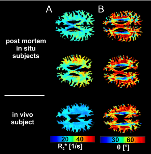
Post mortem in situ subjects had an average brain temperature of 10.9 ± 4.3° Celsius prior to the MRI scan. Overall, higher R2* values are found in post mortem subjects compared to in vivo conditions, while the fiber angle maps appear similar (see figure 1). Global white matter R2* was approximately 28% higher (p < 0.001) and the mean fiber angle θ was approximately 4% higher (p = 0.013) in post mortem (R2* =30.08 ± 2.31 s-1, θ=59.90 ± 1.22 °) compared to in vivo (R2*=23.48 ± 1.39 s-1, θ=57.58 ± 1.19 °). Furthermore, the results show that R2* increases with increasing fiber angle for both in vivo and post mortem (see figure 2). The orientation dependent contribution was lower post mortem (normalized: a0= 1.057, a1= -0.128, a2= 0.008) compared to in vivo examinations (normalized: a0= 1.120, a1= -0.321, a2= 0.091).Discussion
Although diffusion is strongly reduced post mortem 7, the white matter fiber angle could successfully be determined using DTI. Additionally, the decreased orientation dependency of R2* in post mortem compared to in vivo subjects may occur due to the decreased post mortem brain temperature leading to lower diffusion processes.8,9 Moreover, the post-mortem decomposition of the brain tissue may further influence the orientation sensitivity of R2* of WM fibers.Conclusion
In conclusion, the observed data revealed a decreased R2* fiber orientation dependency in post mortem in situ cases compared to in vivo conditions. This might be caused mainly by the lower brain temperature leading to reduced diffusion as well as by white matter tissue decomposition. Although diffusion is reduced post mortem, the white matter fiber orientation can be successfully estimated using DTI in post mortem in situ examinations.Acknowledgements
No acknowledgement found.References
1. Bender B, Klose U. The in vivo influence of white matter fiber orientation towards B0 on T2* in the human brain. NMR Biomed. 2010;23:1071–1076 doi: 10.1002/nbm.1534.
2. Denk C, Torres EH, Mackay A, Rauscher A. The influence of white matter fibre orientation on MR signal phase and decay. NMR Biomed. 2011;24:246–252 doi: 10.1002/nbm.1581.
3. Kor D, Birkl C, Ropele S, et al. The role of iron and myelin in orientation dependent R2* of white matter. NMR Biomed. 2019;32 doi: 10.1002/nbm.4092.
4. Weber AM, Zhang Y, Kames C, Rauscher A. Myelin water imaging and R2* mapping in neonates: Investigating R2* dependence on myelin and fibre orientation in whole brain white matter. NMR Biomed. 2020;33 doi: 10.1002/nbm.4222.
5. Wharton S, Bowtell R. Gradient echo based fiber orientation mapping using R2* and frequency difference measurements. Neuroimage 2013;83:1011–1023 doi: 10.1016/j.neuroimage.2013.07.054.
6. Lee J, Nam Y, Choi JY, Kim EY, Oh SH, Kim DH. Mechanisms of T2* anisotropy and gradient echo myelin water imaging. NMR Biomed. 2017;30 doi: 10.1002/nbm.3513.
7. Scheurer E, Lovblad KO, Kreis R, et al. Forensic application of postmortem diffusion-weighted and diffusion tensor MR imaging of the human brain in situ. AJNR Am J Neuroradiol 2011;32(8):1518-1524.
8. Jones DK, Horsfield MA, Simmons A. Optimal strategies for measuring diffusion in anisotropic systems by magnetic resonance imaging. Magn Reson Med. 1999 Sep;42(3):515-25. PMID: 10467296.]
9. Hernández-Torres E, Kassner N, Forkert ND, et al. Anisotropic cerebral vascular architecture causes orientation dependency in cerebral blood flow and volume measured with dynamic susceptibility contrast magnetic resonance imaging. J. Cereb. Blood Flow Metab. 2017;37:1108–1119 doi: 10.1177/0271678X16653134.
Figures