0227
Ungated time-resolved cardiac MRI with temporal subspace constraint using SSA-FARY SE1Diagnostic and Interventional Radiology, University Medical Center Göttingen, Göttingen, Germany, 2Partner Site Göttingen, German Centre for Cardiovascular Research (DZHK), Göttingen, Germany
Synopsis
SSA-FARY SE is a simple yet powerful novel method to estimate a suitable temporal basis for subspace reconstructions of time-series data from a potentially very small auto-calibration region. Here, we first describe the general strategy of subspace constraint time-series reconstruction. Then, we show how SSA-FARY SE can be used to estimate a suiteable temporal basis. Finally, we demonstrate its functionality by estimating temporal basis functions from the DC-components of radial spokes in single-slice and Simultaneous Multi-Slice free-breathing cardiac MRI acquisitions.
Introduction and Purpose
Ungated time-resolved cardiac MRI is challenging due to the omnipresent cardiac and respiratory motion, and because of the high computational demands posed by the reconstruction of ungated time-series. To reduce the computational burden while still preserving the rich dynamics of the system, a popular approach is to restrict the reconstruction to a pre-estimated temporal subspace [1,2,3]. Like this, the image reconstruction procedure aims at finding an image coefficient for each basis function. The weighted combination of these coefficients, specified by the basis functions, yields the full time-series. Here, we propose an adapted use of SSA-FARY, originally designed for self-gated MRI [4], for efficient temporal Subspace Estimation (SSA-FARY SE) from small auto-calibration (AC-)regions.Theory
Image ReconstructionThe minimization problem for time-resolved MRI with temporal subspace constraint is given by [1,2]:
$$$x=\underset{x}{\text{argmin}}||y-P\mathcal{F}CTx ||_2^2+\lambda||x||_2^2$$$,
with $$$T$$$ the basis for the temporal subspace, $$$C$$$ the coil sensitivities, $$$\mathcal{F}$$$ the Fourier operator and $$$P$$$ a projection onto the k-space trajectory. $$$x$$$ are the image coefficients to be estimated and $$$y$$$ is the acquired data. The full time-resolved movie can then be composed by $$$Tx$$$. Given a suitable $$$T$$$, the minimization can be performed efficiently by precomputing the sampling kernel as described in [5,6].
Temporal Basis Estimation
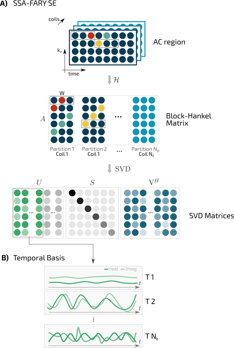
This work focusses on the estimation of the temporal basis $$$T$$$ using SSA-FARY SE. Compared to SSA-FARY for self-gating [4], SSA-FARY SE omits the zero-padding step, thus only two basic mathematical operations need to be performed to determine the temporal basis (FIG_1).
Step 1: Time-Delayed Embedding
A suitable AC-region $$$\boldsymbol{X}$$$ is Hankelized $$$\mathcal{H}$$$ by sliding a window of size $$$W$$$ through the temporal domain of each channel of the AC-region.
$$$\boldsymbol{A}=\mathcal{H}\boldsymbol{X}$$$.
Step 2: Singular Value Decomposition (SVD)
The resulting Block-Hankel matrix $$$A$$$ is decomposed using an SVD
$$$\boldsymbol{A}=USV^H$$$.
The temporal basis $$$T$$$ is obtained by selecting the $$$N_\text{b}$$$ most significant singular vectors of $$$\boldsymbol{A}$$$ given by the columns of $$$U$$$ (FIG_1).
AC-region
The AC-region should contain rich information about the motion during data-acquisition to allow for the estimation of a temporal basis able to accurately describe the temporal process of each pixel. Therefore, a large AC-region, e.g. realized by low-res reconstructions or navigator lines [1], is desirable. This, however, comes at the crucial cost of increased measurement or reconstruction time.
In [4] we have demonstrated that SSA-FARY is capable of detecting motion signals from very small AC-regions through the use of Hankelization (time-delayed embedding). Here, we show that the SSA-FARY approach also improves the estimation of a suitable temporal subspace (SSA-FARY SE).
Methods
In this proof-of-principle free-breathing and ungated cardiac MRI study performed on a Siemens Skyra 3T, we use a radial single-slice (SS, measurement-time=13.2s, base-resolution=2$$$\times$$$192, TR/TE=3.3/1.65ms, bSSFP, flip-angle=40°, tiny-golden-angle=7 [8]) and a radial Simultaneous Multi-Slice (SMS, measurement-time=17.5s, 3 slices, base-resolution=2$$$\times$$$192, TR/TE=2.9/1.79ms, FLASH, flip-angle=12°, tiny-golden-angle=7) acquisition. We furthermore combine 15 or 3$$$\times$$$5 spokes to a frame, which yields the temporal resolution 20.2fps and 23.0fsp, respectively. We make up the AC-region from the DC-component of each spoke, similar to [G], and average the components for each frame. The temporal basis is estimated using SSA-FARY SE with window-size $$$W=21$$$, which covers approximately 1s in both cases. For comparison, we estimate a temporal basis using PCA, which corresponds to SSA-FARY SE with $$$W=1$$$ [1,4], for the SS data. Note, that due to the omitted zero-padding in SSA-FARY SE, the estimated temporal basis is shrinked by $$$W-1$$$ samples, hence about 1s of data are discarded. For better comparison, the PCA-basis was cropped accordingly. All computation was performed with BART [8], including RING gradient-delay estimation [9] and (SMS-)NLINV coil-sensitivity estimation [10,11].Results
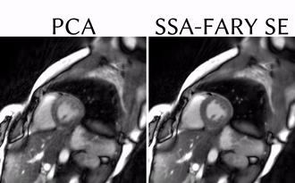
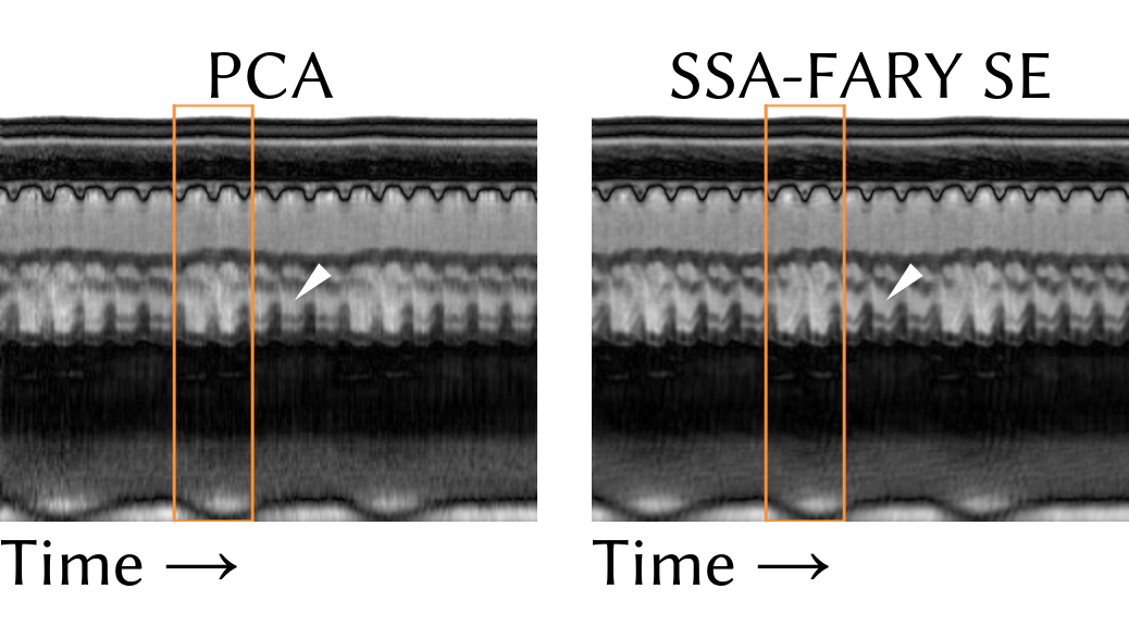
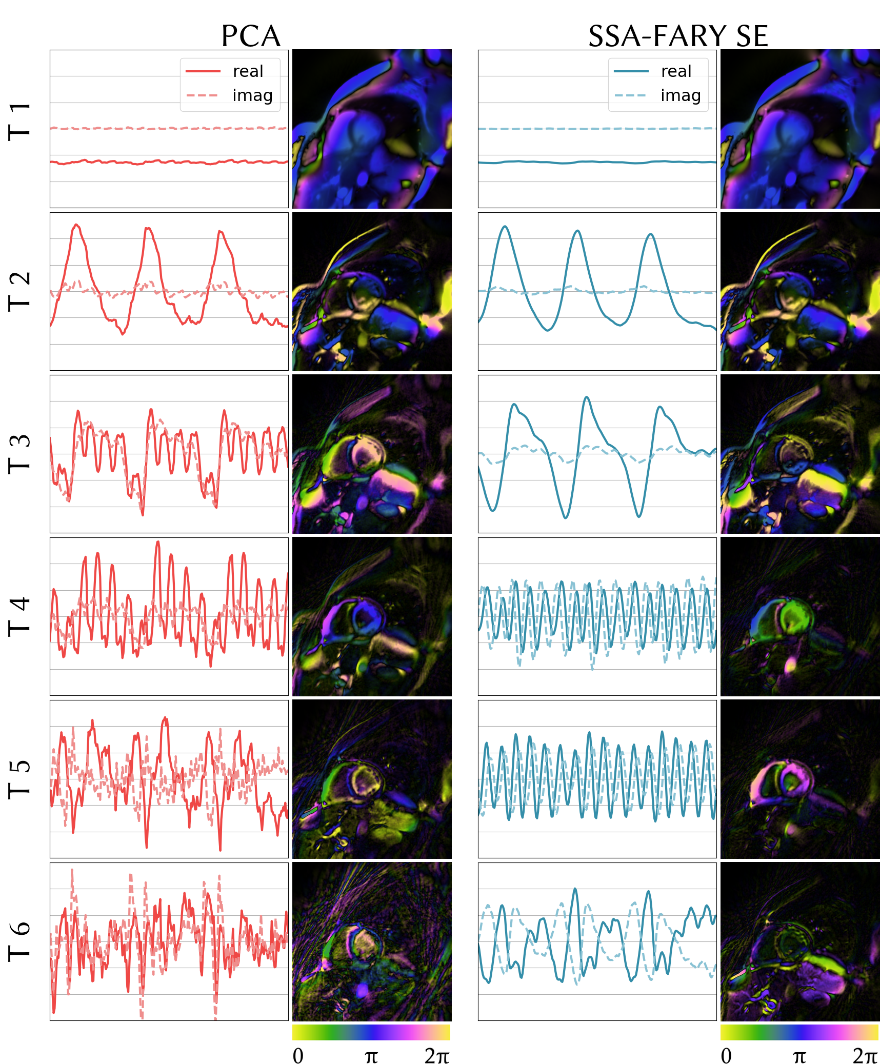
FIG_2 shows a 1.8s snippet of the full SS movie for temporal bases estimated using PCA (left) and SSA-FARY SE (right). The movie reconstructed with the SSA-FARY SE basis looks visually more appealing and can resolve details better than the PCA counterpart (see FIG_3).FIG_4 depicts the six most significant (complex) basis functions estimated with PCA (left) and SSA-FARY SE (right), together with the corresponding complex image coefficients. Note the cleaner basis functions for SSA-FARY SE and the corresponding reduced amount of streakings in e.g. the coefficient for $$$T 6$$$.
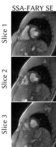
FIG_5 shows a 1.8s snippet of the full SMS movie for three time-consistent slices.
Discussion and Outlook
SSA-FARY SE estimates a suitable temporal subspace for free-breathing cardiac MRI from the center of k-space (DC-component of radial spokes), yielding high-quality movies. Initial results (not shown) furthermore hint that a subspace rank of $$$N_\text{b}=30$$$ yields good quality and that the outcome is relatively insensitive to the choice of the window-size $$$W$$$. In contrast to SSA-FARY for self-gating, the AC region is not zero-padded, complex-valued and $$$W$$$ covers only about one (instead of three) seconds, which ensures a reasonably large bandwidth for the basis-functions [4]. Future work will evaluate the limitations of the method, study the influence of $$$W$$$ and $$$N_\text{b}$$$ on the image quality and investigate advanced regularization strategies [3].Acknowledgements
This work was supported by the German Centre for Cardiovascular Research (DZHK) and funded by the German Research Foundation (DFG) under Grant UE 189/1-1 and in part by NIH under grant U24EB029240.References
[1] Z. Liang "Spatiotemporal imaging with partially separable functions." IEEE Biomed. Imag., vol. 4, pp. 988-991, 2007.
[2] S. Poddar et al. "Free-breathing cardiac MRI using bandlimited manifold modelling." arXiv:1802.08909, (2018).
[3] A. Christodoulou et al. "High-resolution cardiac MRI using partially separable functions and weighted spatial smoothness regularization." In Proc. IEEE Eng. Med. Biol. Soc., pp. 871-874, 2010.
[4] S. Rosenzweig, et al. "Cardiac and Respiratory Self-Gating in Radial MRI Using an Adapted Singular Spectrum Analysis (SSA-FARY)." IEEE Trans. Med. Imag., vol. 39, no. 10, pp. 3029-3041, 2020.
[5] M. Mani et al. "Fast iterative algorithm for the reconstruction of multishot non-cartesian diffusion data." Magn. Reson. Med. 2015, vol. 74, pp. 1086–1094, 2015.
[6] J. Tamir et al. "T2 shuffling: sharp, multicontrast, volumetric fast spin‐echo imaging." Magn. Reson. Med., vol 77, pp. 180-195, 2017.
[7] S. Wundrak et al. "Golden ratio sparse MRI using tiny golden angles." Magn. Reson. Med., vol 75, pp. 2372-2378, 2016.
[8] M. Uecker et al. "Berkeley advanced reconstruction toolbox." In Proc. Intl. Soc. Mag. Reson. Med., vol. 23, p. 2486, 2015.
[9] S. Rosenzweig et al. “Simple auto-calibrated gradient delay estimation from few spokes using radial intersections (RING).” Magn. Reson. Med., vol. 81, no. 3, pp. 1898–1906, 2019.
[10] M. Uecker et al. “Nonlinear inverse reconstruction for real-time MRI of the human heart using undersampled radial FLASH.” Magn. Reson. Med., vol. 63, no. 6, p. 1456–1462, 2010.
[11] S. Rosenzweig et al. “Simultaneous Multi-Slice Real-Time Imaging with Radial Multi-Band FLASH and Nonlinear Inverse Reconstruction.” In Proc. Intl. Soc. Mag. Reson. Med., vol. 24, p. 0518, 2017.
Figures